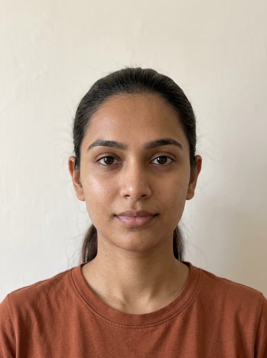
What is canthal tilt?
Canthal tilt is the angle between the inner and outer corners of an eye relative to a horizontal reference line. If the outer corner sits higher, the tilt is positive; if it sits lower, it’s negative; if the corners are about level, it’s neutral.
The inner corner, next to the nose, is the medial canthus. The outer corner is the lateral canthus. In clinical papers, you may see canthal tilt described as the slant or inclination of the palpebral fissure—the opening between the eyelids.
This free test uses Google’s MediaPipe Face Landmarker to estimate each eye’s angle from a photo. You get both angles, their average, and close-ups showing the measurement lines. The results describe this photo, not a fixed measurement of your anatomy. There’s no attractiveness score or target angle.
How to get a more consistent measurement
- Keep your head level and the camera at eye height. Avoid tilting your chin up or down.
- Face the camera straight on and look into the lens. Avoid turning your head to one side.
- Use a relaxed, neutral expression. Don’t smile, squint, or open your eyes unusually wide.
- Use soft, even light from in front of you, so neither eye corner is in shadow.
- Remove glasses and keep hair away from your eyes. A photo without eye makeup makes it easier to see the corners.
- Hold the camera at least an arm’s length away, or ask someone else to take the photo.
Check the corner dots in your result before interpreting the numbers. If repeated photos give different results, check head position and expression first. Similar results across photos can show consistency, but do not establish clinical accuracy.
Reading your results
The large number is the average of the two estimated eye angles. Each eye also has its own angle and label: positive, neutral, or negative.
The close-ups show each eye with the reference line leveled. The blue dots mark the eye corners detected by the model. If a dot misses the corner—for example, because of shadow, mascara, or a squint—the angle for that eye won’t be reliable.
Outer corner higher or lower estimates the height difference between the corners in millimeters. The tool sets the scale using an assumed iris diameter of 11.7 mm. Your actual iris size may differ, so this is an approximation.
Your angle on the scale shows the average as a blue dot and each eye as a gold ring. The scale runs from −12° to +12°; values outside that range appear at the nearest end. It is a display scale, not a normal range.
Estimated difference compares the two eye angles in this photo. Differences of 1° or more are shown numerically; smaller differences are labeled “similar in this photo.” This threshold controls the display, not whether a difference is anatomically meaningful. Small differences may reflect head position or landmark placement. Our face symmetry test explores other differences between the two sides of the face.
Left and right refer to your own sides in an unmirrored photo: your right eye appears on the left. Mirrored selfies swap the labels.
Angles are displayed to one decimal place for readability. That does not mean the tool is accurate to 0.1°. See the limitations before comparing small differences.
Positive, neutral and negative canthal tilt
This tool labels angles from −2° to +2° as neutral, above +2° as positive, and below −2° as negative. The average and each eye are labeled separately.
Neutral on this tool: within ±2° of the pupil line
The ±2° band is our display convention, not a clinical cutoff or a validated accuracy range. A result near either boundary may change categories in another photo.
Clinical morphology terminology uses a different definition: an objectively upslanted or downslanted palpebral fissure has an inclination more than two standard deviations above or below the average for the person’s age. The same terminology notes that the average is slightly upslanting at all ages (Hall et al., 2009).
For an eye opening 30 mm long from corner to corner—an illustrative size close to averages in adult studies—here is how the angle translates to a height difference (Gao et al., 2025; Liu et al., 2023):
| Canthal tilt | Outer corner vs inner corner | This tool’s label |
|---|---|---|
| −6° | 3.1 mm lower | Negative tilt |
| −4° | 2.1 mm lower | Negative tilt |
| −2° | 1.0 mm lower | Neutral tilt |
| 0° | Level | Neutral tilt |
| +2° | 1.0 mm higher | Neutral tilt |
| +4° | 2.1 mm higher | Positive tilt |
| +6° | 3.1 mm higher | Positive tilt |
| +8° | 4.2 mm higher | Positive tilt |
| +10° | 5.2 mm higher | Positive tilt |
At +2°, the outer corner of an eye that size is about 1 mm higher than the inner corner.
Limitations
- Accuracy has not been established for this tool. We have not validated its angles against clinician-marked eye corners. Small differences between eyes or photos may reflect measurement variation rather than a change in anatomy.
- Landmarks are estimates. The model’s eye-corner points may not coincide with the exact points a clinician would mark. Shadows, makeup, and eyelid position can affect placement.
- The reference line matters. We approximate the pupil line using estimated iris centers. A measurement referenced to the inner corners or facial midline can give a different result.
- One photo captures one view. The tool levels sideways tilt, but does not correct for chin position, head turning, perspective, or lens distortion.
- Millimeters depend on an assumed scale. The iris-based conversion is approximate, not a calibrated physical measurement.
- This is not a medical assessment. Results cannot diagnose an eyelid condition or guide surgery.
Published error figures for other systems do not establish this tool’s accuracy. An earlier 468-point face-mesh model had an average landmark-position error of 3.96% of the distance between the eye centers; that is not an eye-angle error in degrees (Kartynnik et al., 2019). A separate study reported 95% limits of agreement of −2.18° to +1.14° for manual measurements minus those from the semiautomated OrbitJ tool. Its fully automated OrbitMap comparison had wider limits, −3.06° to +2.62°. Neither system is the MediaPipe workflow used here (Peterson et al., 2025, Table 3).
Why your result changes between photos
- Chin up or down. Tilting your head forward or back has a strong effect. In a study that photographed 12 women at different head angles, the vertical distance between the corners was about 2.6 mm with the head level, 4.4 mm with the chin tilted 15° down, and close to zero with the chin tilted 15° up (Vaca et al., 2022). Holding the camera above eye level has a similar effect to tilting your chin down.
- Turning your head. When your face is turned, one eye appears shorter, and its angle changes. The photo check rejects photos where the head is clearly turned.
- Expression. Smiling raises the cheeks and lower lids and can change the shape of the outer corner. A squint or a wide-eyed look does the same.
- Makeup and lighting. Eyeliner, mascara and shadows at the corners can shift where the landmarks are placed.
- Mirrored selfies swap your left and right eyes but don’t change the average.
How the angle is measured
The tool estimates 478 facial landmarks, including the eye corners and iris centers. The iris is the colored part of the eye; we use its estimated center as an approximation of the pupil’s position. In this guide, “pupil line” refers to the line joining those two estimated centers.
First, we rotate the landmarks until that line is horizontal. This helps compensate for a slight sideways head tilt. For each eye, we then measure the angle between the reference line and the line from the inner corner to the outer corner, counting upward as positive.
Clinical studies don’t all use the same reference. Standard morphology terminology measures slant relative to a line through the two inner corners, with the head in a neutral position (Hall et al., 2009). Other methods use a horizontal line perpendicular to the facial midline.
We use the estimated pupil line to keep the reference consistent across our tools. The pupil-line and inner-corner methods give the same angles when those two reference lines are parallel—that is, when the inner corners are also level after the pupil line has been leveled.
What is a normal canthal tilt?
Published averages are mostly positive, but they differ between populations and between men and women, so there isn’t one normal value. The studies below used different reference lines and methods, which means their figures aren’t strictly comparable with each other or with this tool:
- Korean adults: reported adult averages of 7.9° in men and 8.8° in women (Park et al., 2008).
- Malaysian South Indian adults aged 18–26: 4.7° in 150 men and 7.1° in 150 women (Packiriswamy et al., 2012).
- Young Black South African women: an average of about 7° in 156 women aged 18–25, ranging from −2.3° to +14.0° (Wilson et al., 2023).
A study using 3D facial models also reported 2D projected angles relative to the inner-corner line. Subtracting its CT_2D values from 180° gives deviations of about 4.9–6.0° in Chinese adults and 0.8–2.4° in Caucasian adults. These are converted 2D values, not the study’s 3D canthal tilt measurements, and do not establish an upward or downward direction (Gao et al., 2025, Table 2).
Within a single group, individual values spread widely. In the South African study they ranged from slightly negative to 14°, showing that slightly negative measurements can occur in healthy young adults. A result from this tool should not be used to decide whether your eyes are healthy.
Does canthal tilt affect attractiveness?
Positive canthal tilt is widely described online as attractive, but the research is mixed and depends on the face and the people rating it:
- Some studies favor a steeper tilt. In an experiment with photos of female faces edited to accentuate what the authors called medial canthal tilt, raters preferred the edited version 93% of the time (Bashour & Geist, 2007). A 3D study found a greater eye-fissure inclination in beauty contest finalists than in a reference group (Sforza et al., 2015), and a study of web images of Black women found a greater tilt in faces rated most attractive (Patel et al., 2026).
- No difference. A study of Korean beauty pageant contestants found no significant difference in eye slant compared with women in the comparison group (Kim et al., 2018).
- Differences between groups. In a study of composite faces, the attractive East Asian composite had a less steep slant than the average one, while the attractive Caucasian and African composites had steeper slants (Rhee et al., 2012). A Korean surgical paper describes cosmetic lateral canthoplasty to lower the outer corner in patients seeking a less upward-slanting eye opening (Kim et al., 2016).
- Photo angle. When women were photographed with the chin tilted down, the measured tilt increased and the photos were rated more attractive (Vaca et al., 2022). Part of what people read as eye shape in a photo may come from how the photo was taken.
In online looksmaxxing communities, a positive canthal tilt is often treated as a goal. A 2026 article in a plastic surgery journal lists it among facial measurements that these communities may misapply, noting that they may not account for ethnic and gender differences (Maini & Janis, 2026). “Hunter eyes,” a popular term in these communities, is not a clinical term.
The AI attractiveness test includes canthal tilt in its eye-area component, using +4° as a reference with a tolerance of 4°; the eye area as a whole carries 15% of that score. We chose that reference ourselves rather than taking it from a study, and this page doesn’t use it.
Canthal tilt, age and eyelid health
Studies disagree about whether the outer corner drops with age. Liu and colleagues found that the outer corner sat lower relative to the inner corner in Caucasian adults aged 61–88 than in those aged 19–34 (Liu et al., 2023). An earlier study of 320 people aged 10 to 89 found that aging did not affect the position of the outer corner (van den Bosch et al., 1999).
In eyelid surgery, a neutral or negative tilt is one of the findings surgeons record before procedures that tighten or reposition the outer corner of the lower lid. In one series of 146 such procedures, the tilt was neutral before surgery in 49% of cases and negative in 18% (Tepper et al., 2015). In medical genetics, an unusually upslanted or downslanted fissure is one of many features clinicians may record. It is defined against age-specific averages and is not a diagnosis on its own (Hall et al., 2009).
A photo-based angle isn’t designed to assess eyelid health. If you’re concerned about your eyelids, for example if a lower lid seems to be pulling away from the eye, talk to a doctor.
Sources
Guide maintained by Your Face Report. Last updated November 9, 2026.
- Hall, B. D., Graham, J. M., Jr., Cassidy, S. B., & Opitz, J. M. (2009). Elements of morphology: Standard terminology for the periorbital region. American Journal of Medical Genetics Part A, 149A(1), 29–39.
- Park, D. H., Choi, W. S., Yoon, S. H., & Song, C. H. (2008). Anthropometry of Asian eyelids by age. Plastic and Reconstructive Surgery, 121(4), 1405–1413.
- Packiriswamy, V., Kumar, P., & Rao, K. G. (2012). Photogrammetric analysis of palpebral fissure dimensions and its position in Malaysian South Indian ethnic adults by gender. North American Journal of Medical Sciences, 4(10), 458–462.
- Wilson, S. P., Medapati, V., & Segwapa, K. (2023). Facial anthropometric norms of the young Black South African woman. Plastic and Reconstructive Surgery Global Open, 11(4), e4942.
- Gao, T., Guo, Y., Rokohl, A. C., Fan, W., Lin, M., Ju, S., et al. (2025). Racial and sexual differences of eyebrow and eyelid morphology: Three-dimensional analysis in young Caucasian and Chinese populations. Quantitative Imaging in Medicine and Surgery, 15(1), 882–897.
- Liu, J., Rokohl, A. C., Liu, H., Fan, W., Li, S., Hou, X., et al. (2023). Age-related changes of the periocular morphology: A two- and three-dimensional anthropometry study in Caucasians. Graefe’s Archive for Clinical and Experimental Ophthalmology, 261(1), 213–222.
- van den Bosch, W. A., Leenders, I., & Mulder, P. (1999). Topographic anatomy of the eyelids, and the effects of sex and age. British Journal of Ophthalmology, 83(3), 347–352.
- Rhee, S. C., Woo, K. S., & Kwon, B. (2012). Biometric study of eyelid shape and dimensions of different races with references to beauty. Aesthetic Plastic Surgery, 36(5), 1236–1245.
- Bashour, M., & Geist, C. (2007). Is medial canthal tilt a powerful cue for facial attractiveness? Ophthalmic Plastic and Reconstructive Surgery, 23(1), 52–56.
- Sforza, C., Dolci, C., Grandi, G., Tartaglia, G. M., Laino, A., & Ferrario, V. F. (2015). Comparison of soft-tissue orbital morphometry in attractive and normal Italian subjects. The Angle Orthodontist, 85(1), 127–133.
- Kim, Y. C., Kwon, J. G., Kim, S. C., Huh, C. H., Kim, H. J., Oh, T. S., et al. (2018). Comparison of periorbital anthropometry between beauty pageant contestants and ordinary young women with Korean ethnicity: A three-dimensional photogrammetric analysis. Aesthetic Plastic Surgery, 42(2), 479–490.
- Patel, A. A., Garg, S. P., Wan, R., Shah, K. V., Ullrich, P., Adam, T. H., et al. (2026). Periorbital measurements of attractiveness in young Black women: An anthropometric approach. Journal of Craniofacial Surgery, 37(3–4), 644–648.
- Kim, Y. J., Lee, K. H., Choi, H. L., & Jeong, E. C. (2016). Cosmetic lateral canthoplasty: Preserving the lateral canthal angle. Archives of Plastic Surgery, 43(4), 316–320.
- Maini, R. S., & Janis, J. E. (2026). Looksmaxxing and the digital evolution of male beauty standards: Ethical implications in plastic surgery. Plastic and Reconstructive Surgery Global Open, 14(8), e7983.
- Tepper, O. M., Steinbrech, D., Howell, M. H., Jelks, E. B., & Jelks, G. W. (2015). A retrospective review of patients undergoing lateral canthoplasty techniques to manage existing or potential lower eyelid malposition: Identification of seven key preoperative findings. Plastic and Reconstructive Surgery, 136(1), 40–49.
- Vaca, E. E., Bricker, J. T., Mioton, L. M., Fagien, S., & Alghoul, M. S. (2022). The influence of sagittal head tilt on periorbital appearance: Implications for clinical photography and the evaluation of postoperative results. Aesthetic Surgery Journal Open Forum, 4, ojab043.
- Peterson, J. C., Nahass, G. R., Lasalle, C., Bradley, D. C., Wu, D., Zorra, I., et al. (2025). Development and validation of a semiautomated tool for measuring periorbital distances. Ophthalmology Science, 5(6), 100887.
- Kartynnik, Y., Ablavatski, A., Grishchenko, I., & Grundmann, M. (2019). Real-time facial surface geometry from monocular video on mobile GPUs. arXiv:1907.06724.
- Google AI Edge. Face landmark detection guide. Documentation for the 478-landmark model used by this tool.